-
Board-Certified
Fellowship-Trained
Physical Medicine
& Rehabilitation
Select from the following links to learn more about Scoliosis.
Flatback Syndrome | Pediatric Scoliosis | Adult Scoliosis | Surgical Options | Harrington Rod | Lordosis | Kyphosis
Scoliosis and spinal deformity
Overview | Causes | Symptoms | Diagnosis | Treatment | FAQ
Overview
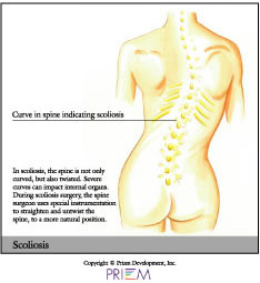
Scoliosis is a disease characterized by an abnormal curvature to the spine, in which the vertebrae twist like a bent corkscrew. In less severe cases, scoliosis may cause the bones to twist slightly, making the hips or ribs appear uneven. When this occurs, the problem is more cosmetic and less of a health risk.
Scoliosis does present a health risk if bones are so
severely twisted that they compress vital organs, or if the spinal
deformity is so severe that spine health and posture is threatened.
If this happens, surgery may be necessary. If left untreated, severe
cases of scoliosis can shorten a person's life span.
[top]
Causes
The exact cause of scoliosis is unknown. Only 1-4 percent of the population
has this condition. It is more common in women than men and most
often affects adolescents between the ages of 10 and 18. A child's
likelihood to develop scoliosis is much higher if their parent or
a sibling has it. Scoliosis can also develop over time in mid- to
late childhood, usually before puberty. In other cases, the disease
is congenital, meaning a person is born with a vertebral abnormality
that causes it.
[top]
Symptoms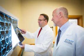
Sometimes, the symptoms of scoliosis are visible. For instance, the
child may have uneven shoulders, chest, hips, shoulder blades, waist,
or a child may have a tendency to lean to one side. In other cases,
there are no visible symptoms. To diagnose a child with scoliosis,
have them touch their toes. If either one or both shoulder blades
are prominent, the waist is shifted or ribs are uneven, scoliosis
may be present. For a child or teenager, your pediatrician often
screens for scoliosis. There are school screening programs as well.
[top]
Diagnosis
Outlined below are some of the diagnostic tools that your physician may use to gain insight into your condition and determine the best treatment plan for your condition.
Medical history: Conducting a detailed medical history helps the doctor better understand the possible causes of your back and neck pain which can help outline the most appropriate treatment.
Physical exam: During the physical exam, your physician will try to pinpoint the source of pain. Simple tests for flexibility and muscle strength may also be conducted.
 X-rays are usually the first step in diagnostic testing
methods. X-rays show bones and the space between bones. They are
of limited value, however, since they do not show muscles and ligaments.
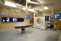
MRI (magnetic resonance imaging) uses a magnetic field and radio waves to generate highly detailed pictures of the inside of your body. Since X-rays only show bones, MRIs are needed to visualize soft tissues like discs in the spine. This type of imaging is very safe and usually pain-free.
CT scan/myelogram: A CT scan is similar to an MRI in that it provides diagnostic information about the internal structures of the spine. A myelogram is used to diagnose a bulging disc, tumor, or changes in the bones surrounding the spinal cord or nerves. A local anesthetic is injected into the low back to numb the area. A lumbar puncture (spinal tap) is then performed. A dye is injected into the spinal canal to reveal where problems lie.
Electrodiagnostics: Electrical testing of the nerves and spinal cord may be performed as part of a diagnostic workup. These tests, called electromyography (EMG) or somato sensory evoked potentials (SSEP), assist your doctor in understanding how your nerves or spinal cord are affected by your condition.
Bone scan: Bone imaging is used to detect infection, malignancy, fractures and arthritis in any part of the skeleton. Bone scans are also used for finding lesions for biopsy or excision.
 Discography is used to determine the internal structure
of a disc. It is performed by using a local anesthetic and injecting
a dye into the disc under X-ray guidance. An X-ray and CT scan are
performed to view the disc composition to determine if its structure
is normal or abnormal. In addition to the disc appearance, your doctor
will note any pain associated with this injection. The benefit of
a discogram is that it enables the physician to confirm the disc
level that is causing your pain. This ensures that surgery will be
more successful and reduces the risk of operating on the wrong disc.
Injections: Pain-relieving injections can relieve back pain and give the physician important information about your problem, as well as provide a bridge therapy.
[top]
Treatment
There are roughly three tiers of treatment for adolescent scoliosis. General scoliosis treatment options include observation, bracing, and if the curve is large and progressive, surgery. Patients with pain and function issues can be treated with therapy, as well as physiatry (physical medicine and rehabilitation physician-supervised programs). Sometimes, shoe inserts (orthotics) are prescribed for those whose legs are uneven.
For adults, the emphasis is on function and movement. Bracing is used only as a temporary pain relief measure; it cannot correct the curve in an adult. Treatment focuses on medications and physical therapy. If other problems exist that are caused by the scoliosis (sacroiliac dysfunction, flatback, spinal stenosis, nerve root pinching), there are many non-operative treatments for each of these issues.
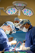
 Surgery may be required in order to correct the spinal
curve. Surgery is usually only recommended for large, progressive curves
or in those patients who have nerve pain that steadily worsens. These
surgeries can be extremely complicated, and a person should invest
a great deal of time in selecting a spine surgeon who subspecializes
in using the most current (fourth generation) corrective techniques.
As with any spine surgery, finding a doctor with experience in this
specific type of surgery is key.
As with any disease, the sooner the problem is discovered, the more treatment options there are available to arrest the progress of the condition.
Click here to visit the website of Axial Biotech and learn more about current spinal disease research.
[top]
FAQs
How can I tell if I have scoliosis?
Your doctor will take X-rays of your spine which will reveal whether or not scoliosis is present as well as how severe it may be.
When is scoliosis considered dangerous to my health?
Scoliosis can be life-threatening when bones are so severely twisted that they compress vital organs. Surgery is most likely the best option in such cases. If left untreated, severe cases of scoliosis can shorten a person's life span.
What are some of the nonsurgical ways to treat scoliosis?
There are some nonsurgical ways to treat scoliosis such as physical
therapy, exercise, bracing, shoe inserts and medication. However, only
a spine surgeon can determine if any of these options might apply to
you.
[top]
Physician Biographies
-
 Karl Boellert, MD
 -
 Mathew Gowans, MD
- Board-Certified
Physical Medicine
& Rehabilitation
- Board-Certified
-
 Michael P.B. Kilburn, MD
-
Board-Certified
Fellowship-Trained
Spinal Neurosurgeon
-
Board-Certified
-
 Sumeer Lal, MD
-
Board-Certified
Fellowship-Trained
Spinal Neurosurgeon
-
Board-Certified
-
 John Cole, MD
-
Board-Certified
Fellowship-Trained
Spinal Neurosurgeon
-
Board-Certified
Back to Life Journal

Home Remedy Book

![]() Website Design & Educational Content © Copyright 2023 Prizm Development, Inc.
Developing Centers of Excellence for Better Healthcare.
Website Design & Educational Content © Copyright 2023 Prizm Development, Inc.
Developing Centers of Excellence for Better Healthcare.
Prizm is the most experienced developer of spine and orthopedic centers in the U.S. with content-rich educational web sites for physicians.
